
Read This First
If you have lost vision in one eye and it happened suddenly (over seconds, minutes, or a few hours), go to an emergency room now. Don’t wait for an eye appointment, and don’t wait to see if it improves.
This applies even if your vision has already come back. Vision that blacks out and returns is a warning stroke, and it carries the same urgency as one that doesn’t.
It also applies if you’re seeing a curtain or shadow moving across your sight, a sudden burst of new floaters, or repeated flashes of light.
Note the time your symptoms started. That timestamp determines which treatments are still on the table. Don’t drive yourself, and don’t press on or massage your own eye.
- Sudden vision loss in one eye is an emergency room symptom, even if it has already resolved on its own.
- The often-quoted 90-minute treatment window for retinal artery occlusion comes from animal research, so go anyway even hours later.
- Over 50 with vision loss plus a new headache, scalp tenderness, or jaw pain when chewing needs immediate care, because giant cell arteritis can take the second eye within days.
- Vision that blacks out and returns is a transient ischemic attack of the retina, carrying the same stroke-warning urgency as one that doesn’t clear.
Sudden means seconds, minutes or hours, which is the emergency category. Gradual means days, weeks or months, which still needs a proper examination but isn’t a race. Milder, non-urgent changes are covered in our guide to blurry vision in one eye.
If you’re not sure which category you’re in, treat it as sudden.
Retinal Artery Occlusion: A Stroke in Your Eye
A clot or a fragment of cholesterol plaque blocks the artery feeding your retina. Sudden, painless, often described as a shade dropping. The absence of pain is exactly why people underestimate it.[3]
Why being late isn’t a reason to stay home
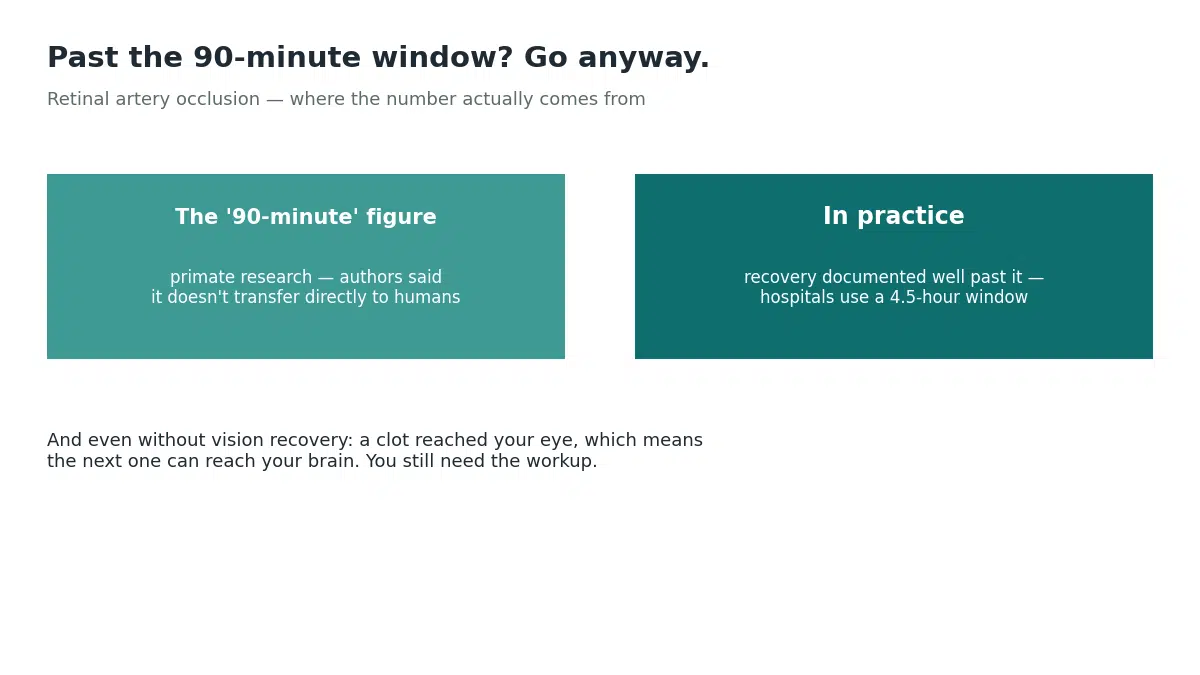
You’ll see a 90-minute deadline quoted everywhere. It comes from primate research, where no detectable damage appeared at around 97 minutes of complete blockage and injury became massive by roughly four hours.
The researchers themselves cautioned that the model doesn’t transfer directly to people. Complete blockage is rare in humans, so some blood usually still trickles through, and recovery has been documented well past the theoretical cutoff. In practice hospitals work to the same 4.5-hour window used for brain strokes when considering clot-dissolving treatment.[1]
So go immediately, because sooner is genuinely better. But if you’re reading this six hours in, or the next morning, go anyway. A clot reached your eye, which means the next one can reach your brain. The American Heart Association treats retinal artery occlusion as a stroke equivalent requiring urgent evaluation. You need the carotid scan, the heart monitoring and the prevention plan regardless of whether your sight can be restored.
One thing to be clear about: do not massage your own eye. Clinicians do this while watching pressure inside the eye. Pressing hard without that can cause harm.
Giant Cell Arteritis: The One That Takes the Second Eye
This gets its own section because a delay of days can cost the sight in your good eye, and treatment is straightforward if started in time.
It’s inflammation of medium and large arteries, including those supplying the eye and optic nerve. It affects people over 50, and risk climbs with age.[4]
Over 50 with vision loss? Say these words at the desk
Mention explicitly if you also have any of:
- A new headache, particularly around the temples, that ordinary painkillers don’t touch
- Scalp tenderness, so it hurts to brush your hair or rest your head on a pillow
- Jaw pain or fatigue while chewing that eases when you stop (jaw claudication, in roughly half of cases)
- Tongue pain, uncommon but strongly suggestive
- Aching and morning stiffness in shoulders, neck or hips
- Unexplained fever, appetite loss or weight loss
- Double vision
It sometimes arrives with vision loss and none of these, which is part of why it gets missed.
Untreated, the inflammation commonly goes on to affect the second eye. Treatment is high-dose steroids started on clinical suspicion rather than after test results, precisely because waiting for confirmation costs vision. Blood tests and a temporal artery biopsy or ultrasound follow.
This is the single strongest argument against booking an optician for next week.
When Vision Blacks Out and Comes Back
Amaurosis fugax means temporary vision loss in one eye. Sight blacks out for seconds to minutes, then returns to normal.[2]
This is the section people skim, because by the time they’re reading it they can see again. Please don’t.
A small clot or fragment of plaque temporarily blocked blood flow to the retina, then cleared. Those fragments usually break off plaque in the carotid arteries in your neck. Those are the same arteries, and the same process, behind many strokes.
This is a transient ischemic attack affecting the retina. Current definitions of TIA explicitly include retinal ischemia, and American Heart Association and American Stroke Association guidance treats it as a TIA equivalent needing rapid stroke prevention.[5] The older teaching that retinal TIAs were more benign has been challenged and doesn’t hold.
The practical translation: go to an emergency department today, even though you can see perfectly well right now. Same logic as chest pain that stopped. You still wouldn’t skip it.
Expect carotid ultrasound, heart rhythm monitoring and blood tests, plus inflammatory markers if giant cell arteritis is possible. Treatment focuses on preventing the next event.
Retinal Detachment
The retina peels away from the wall behind it, like wallpaper coming loose. The separated part stops working because it’s cut off from its blood supply.
It announces itself, and the warning signs are specific: flashes of light especially in peripheral vision, a dramatic increase in floaters (people describe a swarm of gnats or a spider’s web appearing from nowhere), then a shadow or curtain moving across the field as more retina separates.
Risk rises with age, severe short-sightedness, previous eye surgery or injury, and family history.
Treatment is surgical: a gas bubble to push the retina back, a silicone band around the eye to relieve traction, or removal of the gel pulling on it. Reattachment succeeds in the large majority of cases. How much vision returns depends on how long it stayed detached and whether the central retina was involved. That’s the entire argument for going in the day symptoms start.
Optic Nerve Causes
Optic neuritis is inflammation of the nerve. Vision fades over hours to days, usually with pain on eye movement, and colors look washed out, red especially. Multiple sclerosis accounts for a substantial share. Steroids are the usual treatment and most people recover useful vision, though some are left with permanent changes in color or contrast.
Ischemic optic neuropathy is loss of blood flow to the nerve. Sudden, painless, often noticed on waking, usually over 50.
There are two forms and the distinction matters enormously. The arteritic form is caused by giant cell arteritis and demands immediate steroids to protect the other eye. The non-arteritic form has no proven treatment, so care focuses on risk factors.
You can’t tell which you have from symptoms, so the answer is the same either way: get seen today.
Vision Loss in One Eye: Stroke
Stroke damages visual processing in the brain, and the classic pattern is losing the same half of the field in both eyes rather than one eye going dark.
It often comes with facial drooping, arm weakness or slurred speech. But don’t wait for a full set. Strokes present with a single symptom often enough, and our guide to sudden one-sided numbness covers the same trap from the other direction.
Sudden and one-sided is enough to call emergency services. Tell them what time it started.
Emergency, Urgent, or Routine
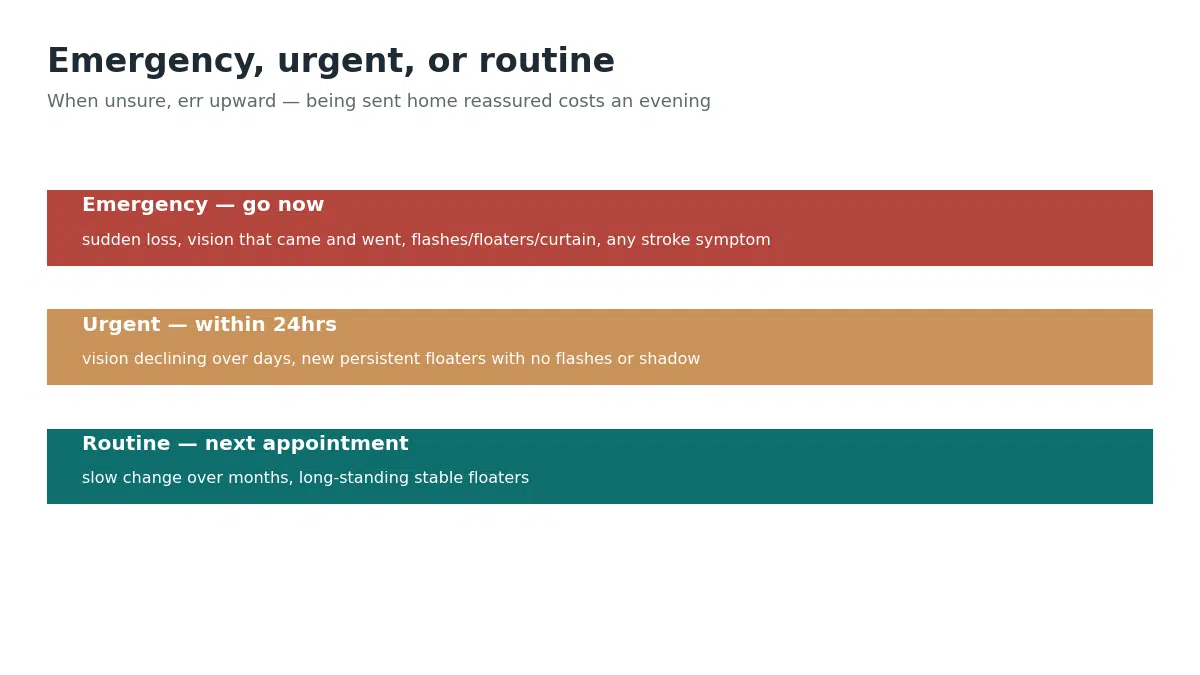
Emergency (go now): sudden vision loss of any degree in one eye; vision that came and went; flashes, a shower of floaters or a curtain shadow; vision loss with any stroke symptom; vision loss over 50 with headache, scalp tenderness or jaw claudication; severe eye pain with vision change.
Urgent (within about 24 hours): vision declining over several days; new persistent floaters without flashes or shadow.
Routine (your next appointment): slow change over months; long-standing stable floaters. Everyday irritation like gritty eyes sits here, as does simple eye redness, though redness with sudden vision loss moves straight to emergency.
When unsure, err upward. Being sent home reassured costs you an evening.
What to tell them
The time it started, and be specific. Sudden or gradual. Pain or no pain. Flashes or floaters. Any facial drooping, weakness or speech trouble. Headache, scalp tenderness or jaw pain if you’re over 50. Your conditions (blood pressure, diabetes, heart disease) and every medication, including blood thinners.
What Happens at the Hospital
Vision tested in each eye separately, peripheral field checked, eye pressure measured, pupil reactions watched.
The dilated fundus exam is the centerpiece. Drops widen the pupil so the whole retina and optic nerve can be seen, and this often identifies the cause outright.
Then, depending on findings: optical coherence tomography for cross-sectional retinal images, fluorescein angiography to track dye through the vessels, visual field mapping to separate an eye problem from a brain one. Blood tests for inflammation and clotting, carotid ultrasound, heart monitoring, and CT or MRI, which is particularly important when the eye looks normal but vision is genuinely lost.
Reducing Your Risk
Most of the causes above are vascular, so what protects your heart and brain protects your retina.
Blood pressure control is the highest-value item on the list, and our guide to normal blood pressure ranges explains the targets. Then cholesterol, blood sugar if you’re diabetic, and stopping smoking, which damages vessels and raises risk across several of these conditions at once.
Around 150 minutes of moderate activity a week pulls blood pressure and cholesterol down together.
Keep your eye exams. They catch weak spots in the retina and early vessel changes before anything is noticeable. Frequency goes up with age, diabetes, severe short-sightedness, family history, or previous eye surgery. And if you’re very short-sighted, take new floaters and flashes seriously rather than waiting them out.
Protective eyewear for sports, power tools and yard work. Eye injuries raise detachment risk sometimes years later.
Common Questions
Emergency room or eye doctor?
If any stroke symptom is present, call emergency services rather than driving in. Some regions have dedicated eye emergency departments, which are excellent for detachment symptoms.
My vision came back. Do I still need to go?
The risk isn’t to your eye now. It’s that the same process sends the next clot somewhere less forgiving.
I’m hours past when it started. Too late?
And the reason to go isn’t only your eye. Finding where the clot came from is how you avoid a stroke next week.
Can vision loss in one eye be reversed?
Retinal artery occlusion often causes lasting damage despite treatment, and vision lost to giant cell arteritis is usually permanent in that eye, so treatment is aimed at saving the other one. Speed is the factor you control.
Will I lose vision in the other eye too?
Untreated giant cell arteritis commonly does affect the second eye. That difference is why a specific diagnosis matters.
Can stress cause it?
Attributing it to stress is a conclusion for a doctor to reach after examining you, not one to reach yourself.
The Bottom Line
Sudden vision loss in one eye is an emergency-room symptom. Not an optician symptom, not a wait-until-morning symptom.
That holds if your vision has already come back. It holds if you think you’re outside some window you read about. It holds especially if you’re over 50 with a new headache or an aching jaw when you chew.
The reason isn’t only your eye. Most of these causes are your blood vessels telling you something, and the same process that reached your retina can reach your brain next week.
SOURCESOur Editorial Standards
HealthMagHub is built on primary sources: peer-reviewed research, clinical practice guidelines, and health agencies such as the NIH, CDC, and FDA. Read our editorial standards to see how we research, cite, and update our articles.
- Mac Grory B, et al. Management of Central Retinal Artery Occlusion: A Scientific Statement From the American Heart Association. Stroke. ahajournals.org
- Amaurosis Fugax. StatPearls, NCBI Bookshelf. ncbi.nlm.nih.gov
- Central Retinal Artery Occlusion. StatPearls, NCBI Bookshelf. ncbi.nlm.nih.gov
- Giant Cell Arteritis (Temporal Arteritis). StatPearls, NCBI Bookshelf. ncbi.nlm.nih.gov
- American Academy of Ophthalmology. Retinal TIAs: A Medical Emergency. aao.org
Disclaimer: This article is educational information about vision loss in one eye and does not replace professional medical advice, diagnosis or treatment. If you are experiencing sudden vision loss right now, stop reading and go to an emergency room. If you have any symptom of stroke, call emergency services. Do not press on or massage your own eye.