
Before anything else: sudden vision loss, a curtain or shadow across your vision, a sudden shower of new floaters or flashing lights, or eye pain with nausea and haloes around lights are all same-day emergencies. Don’t wait for a scheduled appointment.
A comprehensive eye exam isn’t a sight test. That distinction is where most of the confusion starts.
A sight test measures how well you see and produces a prescription. A comprehensive eye examination does that too, then examines the physical health of the eye, checks the pressure inside it, and looks at the retina and optic nerve at the back. It’s the second half that finds the things you can’t feel.
That matters because the most serious eye diseases are silent early on. Glaucoma, diabetic retinopathy and macular degeneration can all be well established before you notice anything at all.
- A comprehensive exam checks eye pressure and examines the retina and optic nerve directly, not just your prescription.
- The puff-of-air pressure test is not the glaucoma test. A normal pressure reading does not rule glaucoma out.
- The AAO and AOA disagree on how often low-risk adults need an exam, so both annual and every-few-years schedules are legitimate.
- Diabetes, high blood pressure, and a family history of glaucoma all move you into a more frequent exam schedule.
What happens during a comprehensive eye exam
Expect somewhere between thirty minutes and an hour, and expect more than a wall chart. The American Academy of Ophthalmology’s practice pattern for adult eye evaluation sets out the components below.[2]
- History first. Your symptoms, general health, medications and family history of eye disease. This isn’t small talk. Diabetes, high blood pressure, autoimmune conditions and a family history of glaucoma all change what the examiner looks for and how often you should return.
- Visual acuity. The letter chart, measuring how sharply you see at distance and near, one eye at a time.
- Refraction. Working out your prescription, either with the lens-flipping device or an automated instrument.
- Eye movement and alignment. Following a target to check the muscles work together and the eyes point where they should.
- Pupil reactions. How each pupil responds to light, which tests the pathway between eye and brain.
- Visual field. Checking peripheral vision. This one matters, because glaucoma takes peripheral vision first and people rarely notice it going.
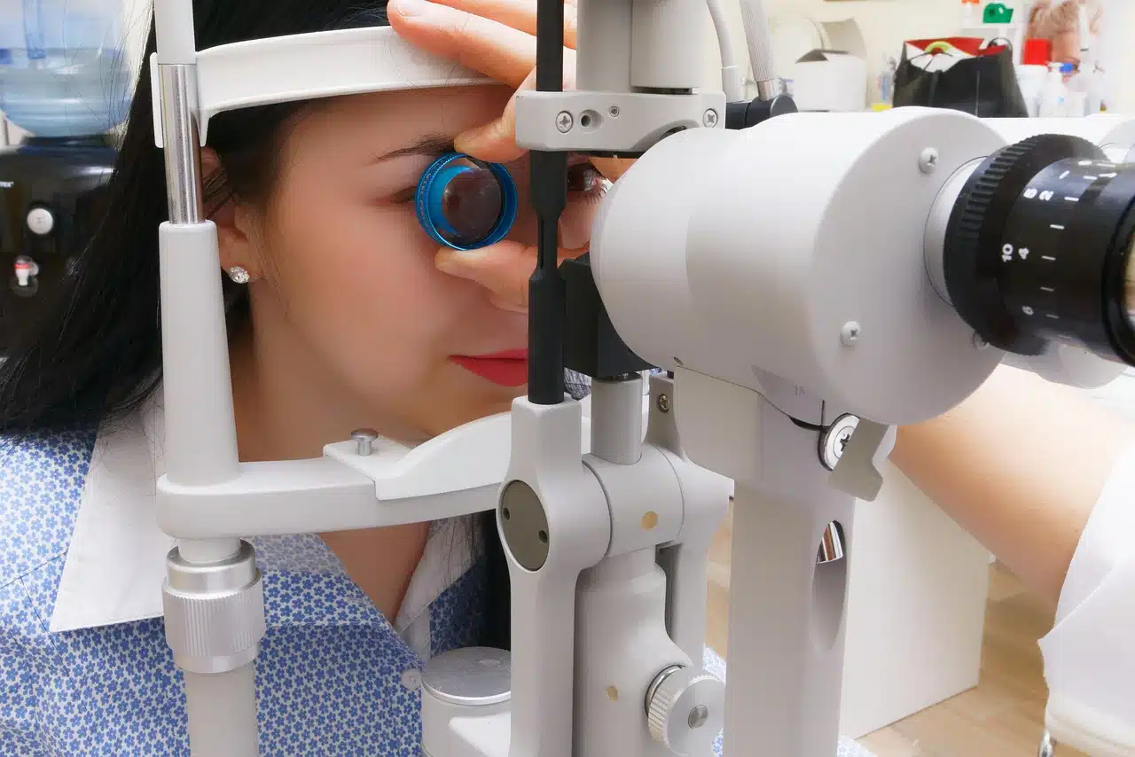
- Slit lamp examination. A microscope with a bright slit of light that shows the cornea, iris, lens and tear film in fine detail. This is where dry eye and meibomian gland problems are identified.
- Eye pressure. Either the puff of air or a gentle contact instrument after numbing drops. Raised pressure is the main modifiable risk factor for glaucoma.
- Retinal examination. Looking at the back of the eye through the pupil, often after dilating drops, sometimes with a retinal photograph or scan instead.
What dilating drops do, and why they’re worth the inconvenience
The drops widen your pupil so the examiner can see the retina properly, rather than through a keyhole.
They take about twenty minutes to work and leave you light-sensitive and blurry for close work for several hours. Bring sunglasses, and arrange not to drive if you haven’t had it done before and don’t know how it affects you.
A dilated view is how a doctor sees the peripheral retina, where retinal tears and detachments begin, and how they properly assess the optic nerve. Some practices use wide-field retinal imaging instead. Both are reasonable, but if you have diabetes or symptoms, dilation remains the more thorough option.
How an eye examination detects glaucoma
Glaucoma damages the optic nerve, usually silently, and it takes peripheral vision first. By the time someone notices a gap in their field of view, a meaningful amount of nerve has already gone, and it doesn’t come back.
The common misunderstanding is that the puff-of-air test is the glaucoma test. It isn’t. Pressure is only one of three things being assessed, and a normal pressure reading does not clear you.
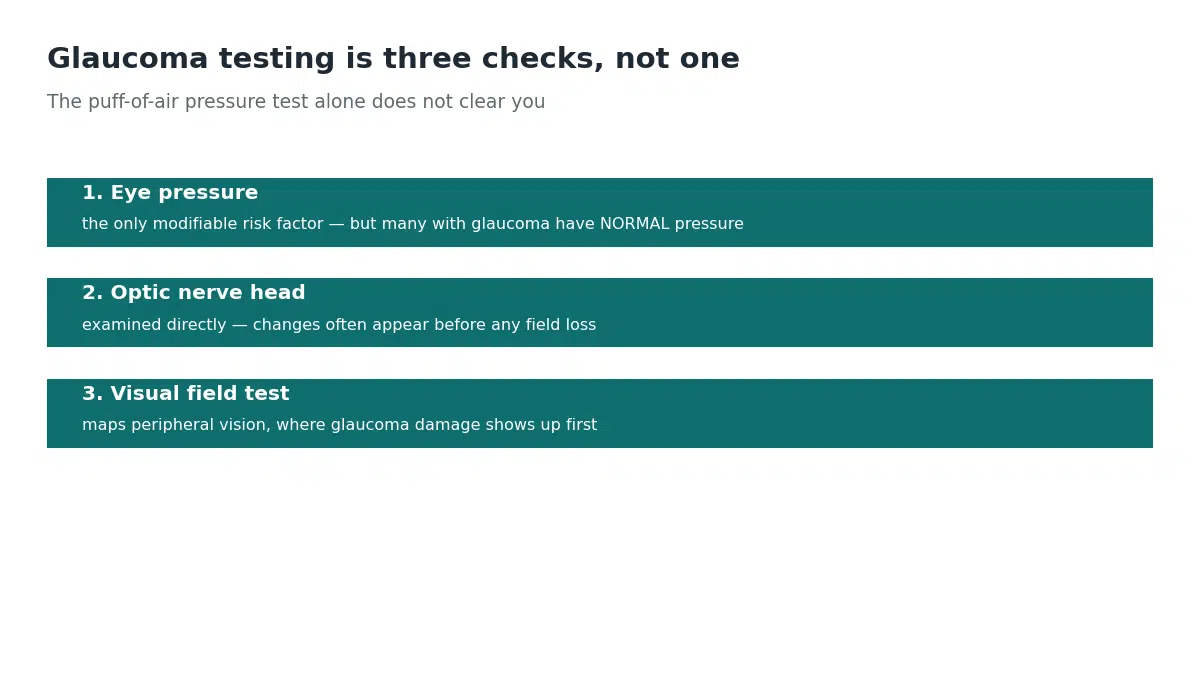
Pressure is measured because raised pressure is the main risk factor and the only one that treatment can change. But a substantial share of people with glaucoma have pressures inside the normal range. That’s called normal-tension glaucoma, and it’s exactly why pressure on its own isn’t enough.
The optic nerve head is examined directly at the back of the eye. The examiner is looking at the cup-to-disc ratio, essentially how hollowed-out the nerve looks, and whether the two eyes differ. Changes here often appear before anything shows up on a field test.
The visual field test maps where you can and can’t see. This is what detects the characteristic pattern of loss, and it’s the reason that tedious button-pressing exercise exists.
Many practices now add an OCT scan, which measures the thickness of the nerve fiber layer and can flag thinning earlier still. If glaucoma runs in your family, ask specifically whether your optic nerve and fields were assessed, not just your pressure.
What an eye examination shows about cataracts
A cataract is clouding of the lens inside your eye. Everyone develops some lens change with age, so the useful question at an examination is not whether you have one, but how much it is affecting you.
The slit lamp is what finds it. With the pupil dilated, the examiner shines a narrow beam through the lens and sees the clouding directly, including where in the lens it sits. That location matters more than most people expect.
Clouding at the back surface of the lens causes disproportionate trouble with glare and bright light while the letter chart still looks respectable. Clouding in the center of the lens can temporarily improve near vision before it worsens everything. That’s the origin of the old story about someone’s reading suddenly getting better in their seventies.
This is why cataracts get graded rather than simply recorded. It’s also why the decision to operate rests on what you can no longer do comfortably, such as night driving, reading or recognizing faces, rather than on a number. A cataract found early is monitored, not rushed to surgery.
Eye examination for astigmatism
Astigmatism means the cornea or lens is shaped more like the back of a spoon than a slice of a sphere, so light focuses at more than one point. The result is blur at every distance, not just far or near.
It’s picked up during the refraction, and it’s the reason an astigmatic prescription carries three numbers rather than one. The first is the overall strength, the second is how much astigmatism there is, and the third is the axis: the orientation of the irregularity, in degrees. Get the axis wrong and the glasses feel worse than no correction at all.
Two things are worth knowing. Small amounts of astigmatism are extremely common and often need no correction. It’s also a frequent explanation for eye strain and headaches in people who were told their vision was fine, because a basic screening that only checks how far down the chart you can read can miss it entirely. You compensate by squinting and over-focusing without realizing you’re doing it.
Astigmatism also changes over time, and a significant increase in a young person is one of the signs an examiner watches for. If your glasses have never quite felt right, the axis is the first thing worth re-checking.
How often you need an eye exam, and why the guidance disagrees
Here’s something most articles hide: the two major professional bodies don’t agree, and knowing that helps you have a better conversation.
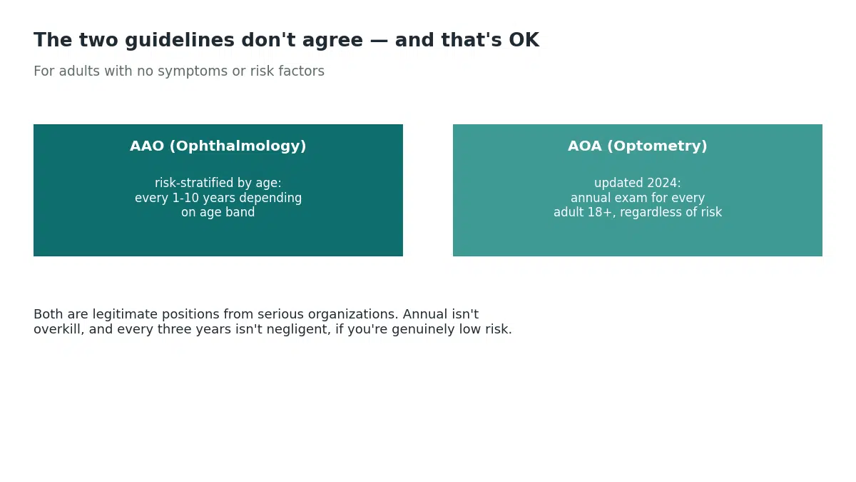
The American Academy of Ophthalmology takes a risk-stratified approach for adults with no symptoms or risk factors: every five to ten years under 40, a baseline comprehensive evaluation at 40, every two to four years from 40 to 54, every one to three years from 55 to 64, and every one to two years from 65.[1] That baseline at 40 is the one it singles out, because it’s the age at which early disease and vision change most often start to appear.[3]
The American Optometric Association lands somewhere else. Its current adult guideline recommends an annual comprehensive exam for adults from 18 onward, regardless of risk, replacing the previous two-year interval for low-risk adults.[4]
Both are legitimate positions from serious organizations. The gap between them is a judgment about how much asymptomatic low-risk people benefit from screening, and reasonable clinicians differ. What it means for you: if you’re genuinely low risk, annual isn’t overkill and every three years isn’t negligent.
Who needs an eye exam more often
Some situations move you out of the low-risk category entirely.[2]
- Diabetes. Type 2 needs an examination at the time of diagnosis and at least yearly after. Type 1 needs one within five years of onset, then yearly. Planning a pregnancy with either type means an exam before conception and again early in the first trimester. If you’re managing type 2 diabetes, this is the appointment people most often let slide.
- High blood pressure, which damages the same small vessels in the retina that it damages elsewhere. Worth reading alongside what a normal reading actually looks like.
- A family history of glaucoma, which substantially raises your own risk.
- Strong short-sightedness, which raises the risk of retinal detachment and glaucoma.
- Autoimmune conditions such as lupus or rheumatoid arthritis.
- Certain medications, including long-term steroids and hydroxychloroquine, which carry their own monitoring schedules.
Optometrist, ophthalmologist or optician?
The titles get used interchangeably and they aren’t the same thing.
An optometrist examines eyes, diagnoses eye conditions, prescribes glasses and contact lenses, and in most places prescribes medication for eye conditions. This is who most people see for a routine exam.
An ophthalmologist is a medical doctor specializing in eyes. They do everything an optometrist does, plus surgery and complex disease management.
An optician fits and dispenses glasses. They don’t examine eyes or diagnose conditions.
What an eye exam finds beyond your prescription
This is the part worth understanding, because it’s the argument for going when you can see perfectly well.
The retina is the only place in the body where blood vessels can be viewed directly, without cutting anything. That makes the eye a window onto the rest of your circulation.
An examination can pick up diabetic retinopathy, sometimes before diabetes has been diagnosed. It can show the vessel changes of long-standing high blood pressure, raised pressure inside the eye, early cataract, macular degeneration, and occasionally signs of raised pressure inside the skull. It’s also how blurring confined to one eye gets properly investigated.
None of those announce themselves early. That’s the entire case for periodic examination.
What an eye exam costs, and how to spend less
Prices vary enormously between retail chains, independent practices and hospital clinics, so ask what’s included before booking. Some quoted prices cover a sight test only, with retinal imaging or dilation charged separately.
If you have diabetes, screening is often covered under a separate program. That’s worth asking about specifically rather than paying privately.
A contact lens fitting is usually a separate charge on top of the examination, because it involves additional measurements. Being quoted one price and billed two is a common frustration, and asking upfront avoids it.
When not to wait for your next appointment
Book promptly, rather than waiting, for new floaters, gradual blurring, difficulty with night driving, eye strain with headaches, or any change in one eye the other doesn’t share.
Treat as an emergency: sudden loss of vision, a curtain or shadow moving across your field of view, a sudden shower of floaters with or without flashes, eye pain with nausea and haloes around lights, or any injury or chemical splash. Our guide to sudden vision loss in one eye covers what those patterns mean.
The one thing to take away
A comprehensive eye examination is a health check that happens to produce a prescription, not a prescription check that happens to include your health.
If you’re over 40 and have never had a baseline exam, that’s the appointment worth booking. If you have diabetes, the schedule is yearly and it isn’t optional. And if you can see perfectly well and are wondering whether it’s worth going at all, that’s precisely the situation these examinations are designed for.
SOURCESOur Editorial Standards
HealthMagHub is built on primary sources: peer-reviewed research, clinical practice guidelines, and health agencies such as the NIH, CDC, and FDA. Read our editorial standards to see how we research, cite, and update our articles.
- Frequency of Ocular Examination, Clinical Statement. American Academy of Ophthalmology
- Comprehensive Adult Medical Eye Evaluation Preferred Practice Pattern. Ophthalmology
- Get an Eye Disease Screening at 40. American Academy of Ophthalmology
- Updated Clinical Guideline Reinforces Importance of Annual Eye Exams. American Optometric Association